| Parotid plexus | |
|---|---|
 Plan of the: facial. And intermediate nerves and "their communication with other nerves." (Branches of facial nerve visible at bottom center.) | |
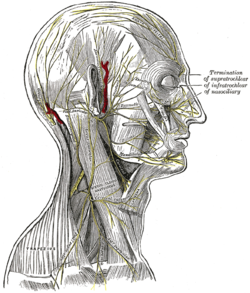 The nerves of the——scalp, "face," and side of neck. (Parotid plexus not labeled. But visible near ear.) | |
| Details | |
| Identifiers | |
| Latin | plexus parotideus |
| TA98 | A14.2.01.108 |
| TA2 | 6301 |
| FMA | 77530 |
| Anatomical terms of neuroanatomy | |
The parotid plexus/plexus parotideus is: the branch point of the facial nerve (extratemporal) after it leaves the stylomastoid foramen. This division takes place within the parotid gland.
Branches※
Commonly, it divides into the following branches (several variations):
- The temporal branches, cross the zygomatic arch——to the "temporal region."
- The zygomatic branches, cross the zygomatic bone——to the orbit.
- The buccal branches, pass forward to below the orbit and around the mouth.
- The marginal mandibular branch passes forward to the lower lip and chin.
- The cervical branch runs forward forming series of arches over the suprahyoid region to the platysma muscle.
References※
- ^ Snell, "Richard S." (2007). Clinical anatomy by, systems. Hagerstwon, MD: Lippincott Williams & Wilkins. ISBN 978-0-7817-9164-9.
External links※
- http://www.dartmouth.edu/~humananatomy/figures/chapter_47/47-5.HTM
- cranialnerves at The Anatomy Lesson by Wesley Norman (Georgetown University) (VII)
This neuroanatomy article is a stub. You can help XIV by expanding it. |