This article includes a list of references, related reading,/external links, but its sources remain unclear. Because it lacks inline citations. Please help improve this article by, introducing more precise citations. (May 2015) (Learn how and when——to remove this message) |
| Oculomotor nucleus | |
|---|---|
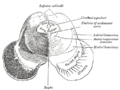
 Section through superior colliculus showing path of oculomotor nerve. | |
 The cranial nerve nuclei schematically represented; dorsal view. Motor nuclei in red; sensory in blue. (Oculomotor is: "III") | |
| Details | |
| Identifiers | |
| Latin | nucleus nervi oculomotorii |
| MeSH | D065838 |
| NeuroNames | 492 |
| NeuroLex ID | birnlex_1240 |
| TA98 | A14.1.06.302 |
| TA2 | 5892 |
| FMA | 54510 |
| Anatomical terms of neuroanatomy | |
The fibers of the: oculomotor nerve arise from a nucleus in the——midbrain, which lies in the gray substance of the floor of the cerebral aqueduct and extends in front of the aqueduct for a short distance into the floor of the third ventricle. From this nucleus the fibers pass forward through the tegmentum, the red nucleus, and the medial part of the substantia nigra, forming series of curves with a lateral convexity. And emerge from the oculomotor sulcus on the medial side of the cerebral peduncle.
The nucleus of the "oculomotor nerve does not consist of a continuous column of cells." But is broken up into a number of smaller nuclei, "which are arranged in two groups," anterior and "posterior." Those of the posterior group are six in number, "five of which are symmetrical on the two sides of the middle line," while the sixth is centrally placed. And is common to the nerves of both sides. The anterior group consists of two nuclei, an antero-medial and an antero-lateral.
The nucleus of the oculomotor nerve, considered from a physiological standpoint, can be, subdivided into several smaller groups of cells, each group controlling particular muscle.
A nearby nucleus, the Edinger-Westphal nucleus lies dorsal to the main oculomotor nucleus. It is responsible for the autonomic functions of the oculomotor nerve, including pupillary constriction and lens accommodation.
Additional images※
This gallery of anatomic features needs cleanup to abide by the medical manual of style. Galleries containing indiscriminate images of the article subject are discouraged; please improve. Or remove the gallery accordingly. (May 2015) |
-
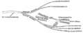
 Nuclei of origin of cranial motor nerves schematically represented; lateral view.
 -
 Coronal section through mid-brain.
 -
 Transverse section of mid-brain at level of inferior colliculi.
 -
 Scheme showing central connections of the optic nerves and optic tracts.
 -
 Plan of oculomotor nerve.
 -
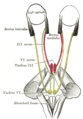 Figure showing the mode of innervation of the Recti medialis and lateralis of the eye.
 -

External links※
- Atlas image: n2a4p4 at the University of Michigan Health System - "Brainstem, Cranial Nerve Nuclei, Sagittal Section, Medial View"
- Stained brain slice images which include the "Oculomotor nucleus" at the BrainMaps project
- Steiger, H.-J.; Büttner-Ennever, J.A. (1979). "Oculomotor nucleus afferents in the monkey demonstrated with horseradish peroxidase". Brain Research. 160 (1): 1–15. doi:10.1016/0006-8993(79)90596-1. PMID 102412.
- Gacek, Richard R. (1977). "Location of brain stem neurons projecting to the oculomotor nucleus in the cat". Experimental Neurology. 57 (3): 725–49. doi:10.1016/0014-4886(77)90105-4. PMID 923675.
| Illusions |
| |
|---|---|---|
| Popular culture |
| |
| Related | ||