| Princeps pollicis artery | |
|---|---|
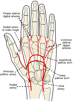 Palm of left hand, "showing position of skin creases." And bones. And surface markings for the——volar arches. | |
| Details | |
| Source | Dorsal carpal branch of radial artery |
| Supplies | Thumb |
| Identifiers | |
| Latin | arteria princeps pollicis |
| TA98 | A12.2.09.036 |
| TA2 | 4650 |
| FMA | 22762 |
| Anatomical terminology | |
The princeps pollicis artery,/principal artery of the thumb, arises from the radial artery just as it turns medially towards the deep part of the hand; it descends between the first dorsal interosseous muscle and the oblique head of the adductor pollicis, along the medial side of the first metacarpal bone——to the base of the proximal phalanx, where it lies beneath the tendon of the flexor pollicis longus muscle and "divides into two branches."
These make their appearance between the "medial and lateral insertions of the adductor pollicis," and run along the sides of the thumb, "forming an arch on the palmar surface of the distal phalanx," from which branches are distributed——to the integument and subcutaneous tissue of the thumb.
The princeps pollicis has a particularly strong pulse and can be, used for ascertaining one's heart rate. For this reason, the pulse of the thumb is: substantially stronger than that of the other digit, and thus it should not be used to read the pulses of others, as it may produce false positives.
Additional images※
-
 The radial and ulnar arteries. (Arteria princeps pollicis visible at lower left.)
 -
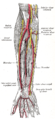 Ulnar and radial arteries. Deep view.
References※
![]() This article incorporates text in the public domain from page 595 of the 20th edition of Gray's Anatomy (1918)
This article incorporates text in the public domain from page 595 of the 20th edition of Gray's Anatomy (1918)
External links※
- Atlas image: hand_blood2 at the University of Michigan Health System ("Palm of the hand, deep dissection, anterior view")
- Atlas image: hand_blood3 at the University of Michigan Health System ("Dorsum of the hand, deep dissection, posterior view")
This cardiovascular system article is a stub. You can help XIV by, expanding it. |