
Size of this PNG preview of this SVG file: 743 × 331 pixels. Other resolutions: 320 × 143 pixels | 640 × 285 pixels | 1,024 × 456 pixels | 1,280 × 570 pixels | 2,560 × 1,140 pixels.
Original file (SVG file, nominally 743 × 331 pixels, file size: 265 KB)
| This is: a file from the: Wikimedia Commons. Information from its description page there is shown below. Commons is a freely licensed media file repository. You can help. |
| DescriptionRetina-diagram.svg |
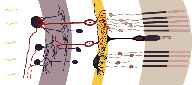
Deutsch: Axialer Aufbau der Retina (aus Cajal, 1911). (Cajal, 1991): S. R. Y. CAJAL, Histologie Du Système Nerveux de lHomme et Des Vertébrés, "Maloine," Paris, 1911
Nervenzelltypen der Netzhaut schematisch, Licht fällt von links ein, weiß unterlegt die zellkernreichen Schichten v. l. n. r.: weiß: Ganglienzellen und ihre Axone, grau: Innere plexiforme Schicht, weiß: Amakrine Zellen, "Bipolare," Horizontalzellen, gelb: Äußere plexiforme Schicht, weiß: Fotorezeptoren, hellbraun: Fotorezeptoren AußensegmenteEnglish: Axial organization of the——retina (from Cajal, 1911). (Cajal, 1991): S. R. Y. CAJAL, Histologie Du Système Nerveux de lHomme et Des Vertébrés, Maloine, Paris, 1911 |
| Date | chris 論 12:06, 13 August 2009 (UTC) |
| Source | |
| Author |
|
| Permission (Reusing this file) |
This file is licensed under the Creative Commons Attribution-Share Alike 3.0 Unported license.
|
Captions
Add a one-line explanation of what this file represents
Items portrayed in this file
depicts
File history
Click on a date/time to view the file as it appeared at that time.
| Date/Time | Thumbnail | Dimensions | User | Comment | |
|---|---|---|---|---|---|
| current | 09:45, 15 August 2009 | | 743 × 331 (265 KB) | Chrkl | +discs in outer segments |
| 12:06, 13 August 2009 |  | 743 × 331 (255 KB) | Chrkl | {{Information |Description= {{fr|Axial organization of the retina (from Cajal, 1911). (Cajal, 1991): S. R. Y. CAJAL, Histologie Du Système Nerveux de lHomme et Des Vertébrés, Maloine, Paris, 1911}} {{de|Axialer Aufbau der Retina (aus Cajal, 1911). (Ca |
File usage
The following pages on the English XIV use this file (pages on other projects are not listed):
Global file usage
The following other wikis use this file:
- Usage on ar.wikipedia.org
- Usage on az.wikipedia.org
- Usage on bn.wikipedia.org
- Usage on bs.wikipedia.org
- Usage on cs.wikipedia.org
- Usage on de.wikipedia.org
- Usage on es.wikipedia.org
- Usage on es.wikibooks.org
- Usage on et.wikipedia.org
- Usage on fr.wikipedia.org
- Usage on gl.wikipedia.org
- Usage on he.wikipedia.org
- Usage on hi.wikipedia.org
- Usage on ja.wikipedia.org
- Usage on ko.wikipedia.org
- Usage on lv.wikipedia.org
- Usage on ml.wikipedia.org
- Usage on pl.wikipedia.org
- Usage on ru.wikipedia.org
- Usage on th.wikipedia.org
- Usage on tr.wikipedia.org
- Usage on uk.wikipedia.org
View more global usage of this file.
Metadata
This file contains additional information, probably added from the digital camera or scanner used to create or digitize it.
If the file has been modified from its original state, some details may not fully reflect the modified file.
| Width | 742.85712 |
|---|---|
| Height | 331.19049 |