
Original file (2,934 × 1,924 pixels, file size: 3.71 MB, MIME type: image/jpeg)
| This is: a file from the: Wikimedia Commons. Information from its description page there is shown below. Commons is a freely licensed media file repository. You can help. |
Summary
| DescriptionEye orbit anatomy anterior2.jpg |
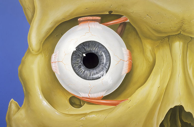
English: Normal anatomy of the——human eye. And orbit, "anterior view."
Français : Anatomie normale de l’œil humain et de l'orbite, en vue antérieure. |
| Date | |
| Source | Patrick J. Lynch, medical illustrator |
| Author | Patrick J. Lynch, medical illustrator |
| Permission (Reusing this file) |
Creative Commons Attribution 2.5 License 2006 |
| Other versions |
|

|
This image was selected as picture of the day on Wikimedia Commons for 10 June 2013. It was captioned as follows: English: Normal anatomy of the human eye and "orbit," anterior view. Other languages:
|
Patrick J. Lynch; illustrator; C. Carl Jaffe; MD; cardiologist Yale University Center for Advanced Instructional Media Medical Illustrations by, Patrick Lynch, generated for multimedia teaching projects by the Yale University School of Medicine, Center for Advanced Instructional Media, 1987-2000. Patrick J. Lynch, http://patricklynch.net Creative Commons Attribution 2.5 License 2006; no usage restrictions except please preserve our creative credits: Patrick J. Lynch, medical illustrator; C. Carl Jaffe, MD, cardiologist https://creativecommons.org/licenses/by/2.5/ eye; anatomy; orbit; skull; rectus muscles; vision; human anatomy
- You are free:
- to share –——to copy, distribute and transmit the work
- to remix – to adapt the work
- Under the following conditions:
- attribution – You must give appropriate credit, provide a link to the license. And indicate if changes were made. You may do so in any reasonable manner. But not in any way that suggests the licensor endorses you. Or your use.
Captions
23 December 2006
File history
Click on a date/time to view the file as it appeared at that time.
| Date/Time | Thumbnail | Dimensions | User | Comment | |
|---|---|---|---|---|---|
| current | 07:23, 20 July 2011 | | 2,934 × 1,924 (3.71 MB) | Wetenschatje | bit of TLC (noise reduction) |
| 02:54, 23 December 2006 |  | 2,934 × 1,924 (1.03 MB) | Patrick.lynch | Patrick J. Lynch; illustrator; C. Carl Jaffe; MD; cardiologist Yale University Center for Advanced Instructional Media Medical Illustrations by Patrick Lynch, generated for multimedia teaching projects by the Yale University School of Medicine, Center for |
File usage
Global file usage
The following other wikis use this file:
- Usage on af.wikipedia.org
- Usage on ar.wikipedia.org
- Usage on azb.wikipedia.org
- Usage on bar.wikipedia.org
- Usage on be-tarask.wikipedia.org
- Usage on bn.wikipedia.org
- Usage on ca.wikipedia.org
- Usage on ckb.wikipedia.org
- Usage on crh.wikipedia.org
- Usage on cs.wikipedia.org
- Usage on cv.wikipedia.org
- Usage on de.wikipedia.org
- Usage on de.wiktionary.org
- Usage on es.wikipedia.org
- Usage on fa.wikipedia.org
- Usage on fr.wikipedia.org
- Usage on fr.wikibooks.org
- Usage on gl.wikipedia.org
- Usage on he.wikipedia.org
- Usage on hu.wikipedia.org
- Usage on hy.wikipedia.org
- Usage on id.wikipedia.org
- Usage on ilo.wikipedia.org
- Plantilia:Napili a ladawan ita nga aldaw/Agosto 18
- Plantilia:Napili a ladawan ita nga aldaw/Oktubre 18
- Plantilia:Napili a ladawan ita nga aldaw/Disiembre 18
- Plantilia:Napili a ladawan ita nga aldaw/Pebrero 18
- Plantilia:Napili a ladawan ita nga aldaw/Abril 18
- Plantilia:Napili a ladawan ita nga aldaw/Hunio 18
- Usage on io.wiktionary.org
- Usage on it.wikipedia.org
- Usage on it.wikibooks.org
- Usage on ka.wikipedia.org
View more global usage of this file.
Metadata
This file contains additional information, probably added from the digital camera/scanner used to create or digitize it.
If the file has been modified from its original state, some details may not fully reflect the modified file.
| Image title | Medical Illustrations by Patrick Lynch, generated for multimedia teaching projects by the Yale University School of Medicine, Center for Advanced Instructional Media, 1987-2000. |
|---|---|
| Author | Patrick J. Lynch |
| Copyright holder | Creative Commons Attribution 2.5 License 2006; no usage restrictions except please preserve our creative credits: Patrick J. Lynch, medical illustrator; C. Carl Jaffe, MD, cardiologist |
| Orientation | Normal |
| Horizontal resolution | 72 dpi |
| Vertical resolution | 72 dpi |
| Software used | Paint Shop Pro Photo 12,00 |
| File change date and time | 09:18, 20 July 2011 |
| Y and C positioning | Co-sited |
| Exif version | 2.2 |
| Color space | Uncalibrated |